+Anonymously helping burn patients and victims....

contact us: asktheburnsurgeon@yahoo.com


Tuesday, April 1, 2014

ASSESSMENT OF BURN SURFACE AREA……


Doc, what happens if I calculate the burn area wrongly? Is it really important?
Arthur A, Canada

As we have seen in previous posts the loss of fluid from the skin surface depends on the degree of burns and the area of the burns. One must evaluate the total burn surface area (TBSA) to calculate the fluid requirements. There are different methods of fluid calculation in burns.
a) Rule of nine
The rule of nine works well in adult patients. In this method the body surface is divided into various parts measuring in nines.
Rule of nine
Each arm - 9% TBSA
Head - 9% TBSA
Anterior thorax - 18% TBSA
Posterior thorax - 18% TBSA
Perineum - 1% TBSA
Each leg - 18% TBSA

Any burn surgeon will tell you that more often the burns are so irregularly placed that accurate calculation becomes difficult in different regions of the body. In such cases a simple trick is to use the palm of the hand as a method of calculation. At any age the palm of the hand is approximately 1% and can be used to measure the burn areas.  One must not forget that it is the patient’s hand that is used for the calculation and not the doctor’s. An approximate size of the palm of the patient is considered and the equivalent burn area is estimated. For e.g. lets say the burn area was 5 palm sizes of the patient over the body and lower limbs. Now we can assume that the patient has about 5% burns.
It should be noted that first degree burns do not produce fluid losses and therefore only 2nd degree burns or more should be used for fluid calculation.
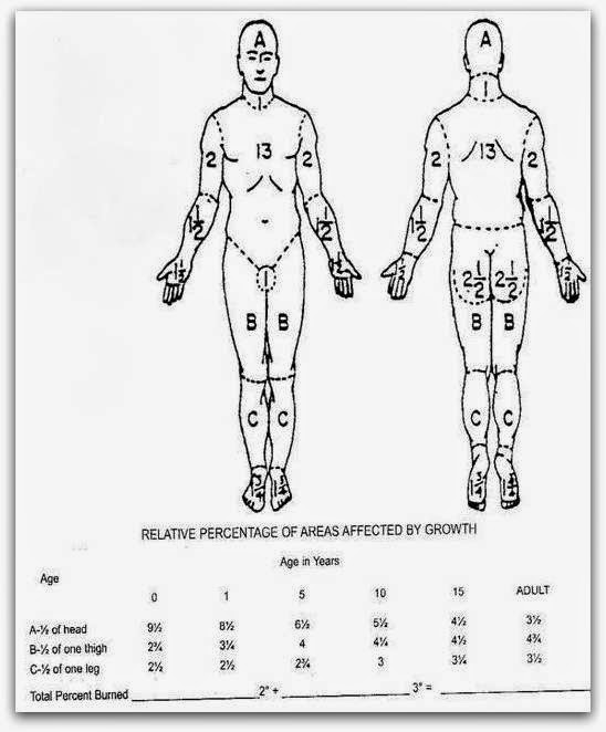
However in children the rule of nine can lead to serious errors as the head and body is larger in TBSA than the limbs and therefore the Lund and Browder charts work out to be more accurate while calculating the fluids to be administered.  The Lund and Browder chart is shown below. 

If the fluid calculation is wrong then the patient will be administered less fluids and this will result in shock or low volume circulatory failure and ultimately may be fatal.  The fluids calculated need to be replaced within a time limit as we shall discuss in the next post. Correct volume replacement and correct timing is what makes the resuscitation of burns patients successful.
(an original initiative in burn care and education from asktheburnsurgeon+)

Sunday, March 30, 2014

How burns affect the tissues.....


Doc, could you please explain the cellular and chemical processes that occur when the tissues are burnt?

Rony v, Goa

A number of inflammatory processes both local (at the site of the burn) as well as systemic (in the rest of the body) take place when burns occur which eventually lead to the shifting of fluid from the vascular compartment to the interstitial spaces. Subsequent to the burn a number of cells like Neutrophils, macrophages, and lymphocytes cross over into the burned tissues and start releasing chemical mediators like histamine, serotonin, prostaglandins, kinins, platelet products and complement components. These chemical substances damage the normal blood capillary barrier which leads to an increase in the permeability of the vessels. Intravascular fluids therefore start to leak from the walls of the vessels and this ultimately leads to a decrease of the circulating intravascular blood volume. A fact that is not commonly recognized is that these processes that occur in the burn tissues also take place in the tissues that have not suffered any burns and therefore one can see edema in areas of the body that have not suffered burns. Thermal injury also ends up damaging the cell wall and collagen fibers which in turn lead to inadequacy of the cell wall transport mechanisms and buildup of sodium and water and eventual death of the cell if the fluid imbalances are not immediately corrected. In minor burns such as 10 percent in children and about 15 percent in adults these fluid balances are well adjusted and tolerated and therefore additional fluid replacements are not needed. However in patients with TBSA higher than the one’s mentioned above intravenous fluid resuscitation is needed.
The capillaries begin to regain their functional integrity and the leak eventually gets controlled, but this often takes more than 8 hours post burn. Crystalloids are usually given in the first 8 hours and one’s the integrity of the capillary wall is regained after 8 hours, colloid fluids are started since they will not leak out. Adding colloids also help to reduce the fluid overload that may result of excess of crystalloid infusions. Burn wounds are composed of three zones- a central zone of coagulation or severe tissue damage, a peripheral zone of hyperemia or vasodilatation, and an  intervening zone of stasis or low blood flow (ischaemia). If the fluid imbalances that occur from the fluid shifts as noted above are not corrected on time then these zones can extend and more tissue damage can result explaining the fact that some superficial burns on admission can end up as deep over period of time. This can also happen when burn wounds get infected.
(an original initiative in burn care education from asktheburnsurgeon)
+